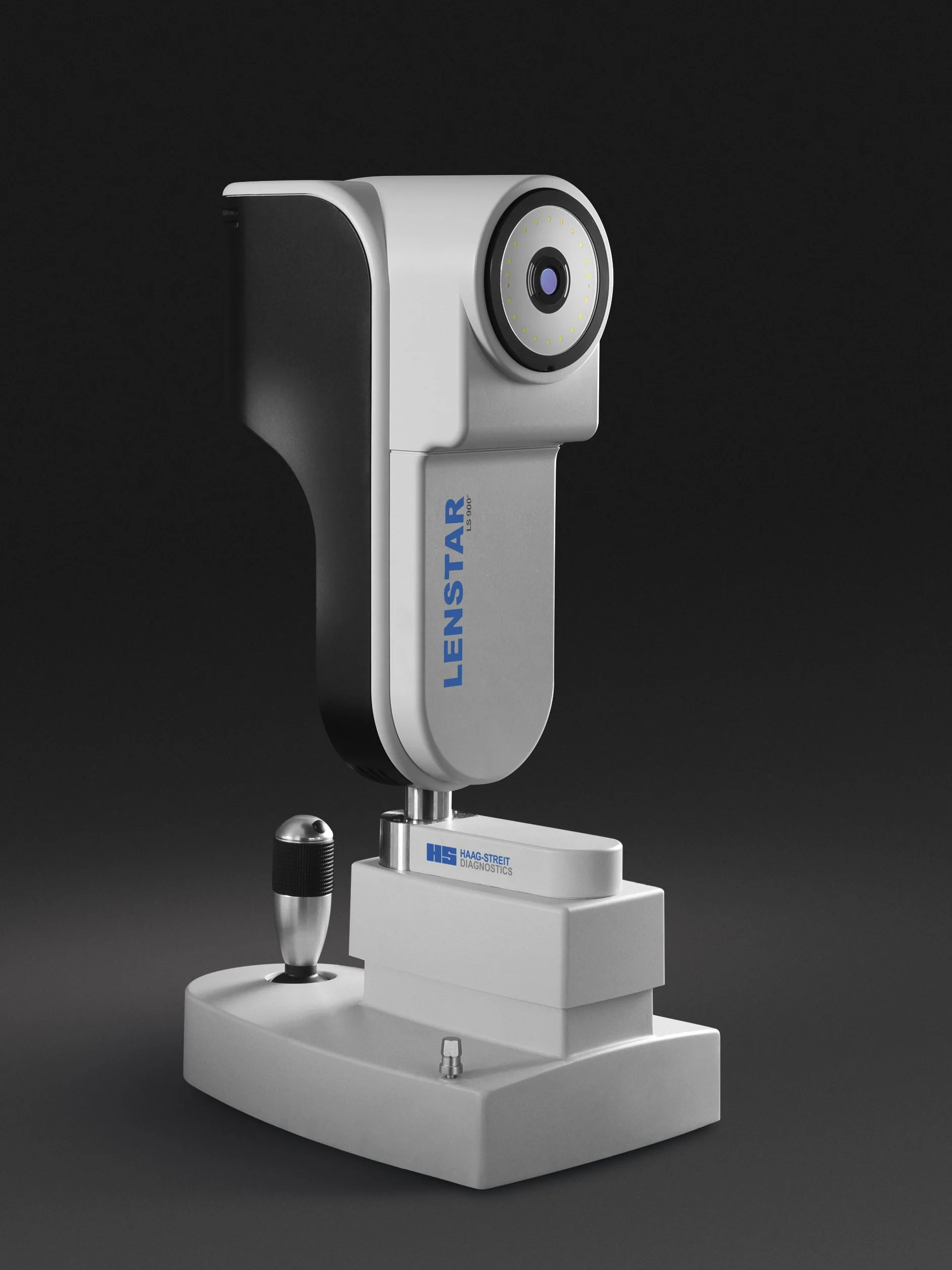
Lenstar Biometry for Myopia Management
The Haag-Streit Lenstar biometer measures the axial length of the eye, a critical measure for assessing the progression of myopia especially in children and adolescents
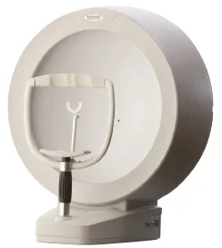
Home » Technology » Automated Perimetry and Visual Fields
Visual acuity, or our ability to see detail, is one of our most important visual functions and is used every day. It determines how well we can read, how well we can see people in the distance and how we manage with detailed visual tasks.
Importantly however, our visual space is not limited to detailed central vision. Peripheral and para-central vision is also critical in our everyday lives. We measure peripheral vision and the extent and completeness of the visual field with an instrument called a perimeter, which is computer-driven and therefore automated.
When doing perimetry, you are required to look directly at the central light of the perimeter bowl. Other lights of varying brightnesses flash, one at a time, anywhere in the central and peripheral fields. You have a response button to press each time you notice one of those lights flashing. It normally takes about ten minutes for each eye to take a detailed field measurement.
Changes to the fields of vision, either the central or the peripheral field, are often a critical sign of serious eye and neurological conditions; these include glaucoma, intracranial tumours and stroke. Visual field testing, also called perimetry, is particularly important in patients at risk of developing glaucoma and those on certain systemic medications.
Learn more about the technology that we use at Collins Street Optometrists

The Haag-Streit Lenstar biometer measures the axial length of the eye, a critical measure for assessing the progression of myopia especially in children and adolescents
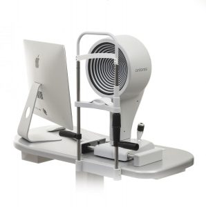
Keratography with the Oculus K5 instrument is an invaluable tool for the assessment, diagnosis and classification of dry eye
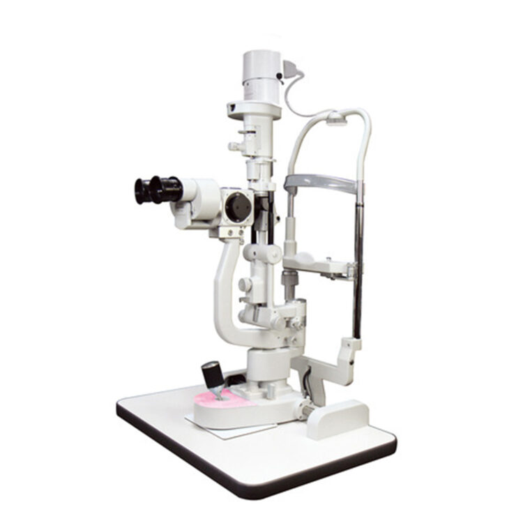
High quality magnification and lighting are essential for examining the front of the eye and detecting subtle changes, which is assisted further by the recording function of digital photography
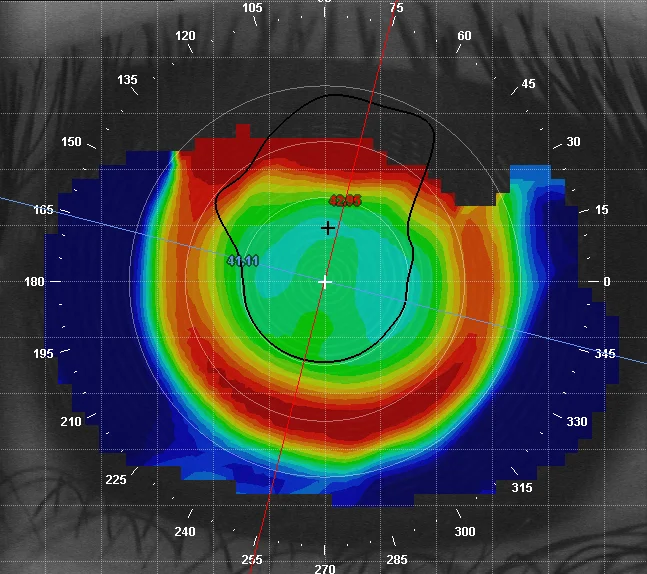
Corneal topography provides a computerised 3-D map of the front surface of the eye, with which we can see the characteristics of its curves and surface irregularities and which is enormously helpful for contact lens fitting
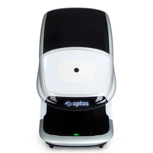
Our Optos Daytona scans the retina with two lasers through a range of 200°, where by comparison, standard digital retinal photography captures only the central 45°
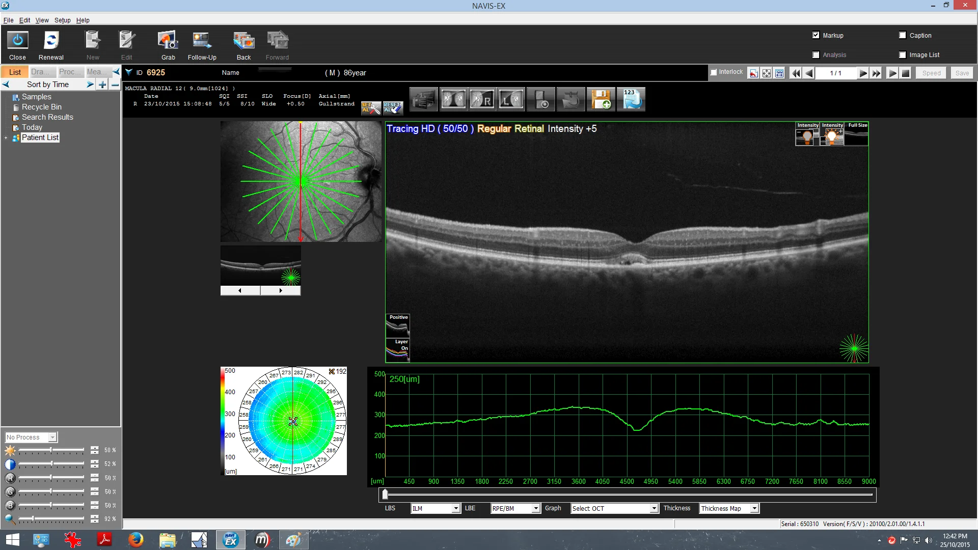
Optical Coherence Tomography is a non-invasive imaging technology used to capture cross-sectional images of the retina and is an essential aid in the diagnosis of retinal disease and glaucoma
Monday - Friday
9AM - 6PM
Saturday - Sunday
CLOSED