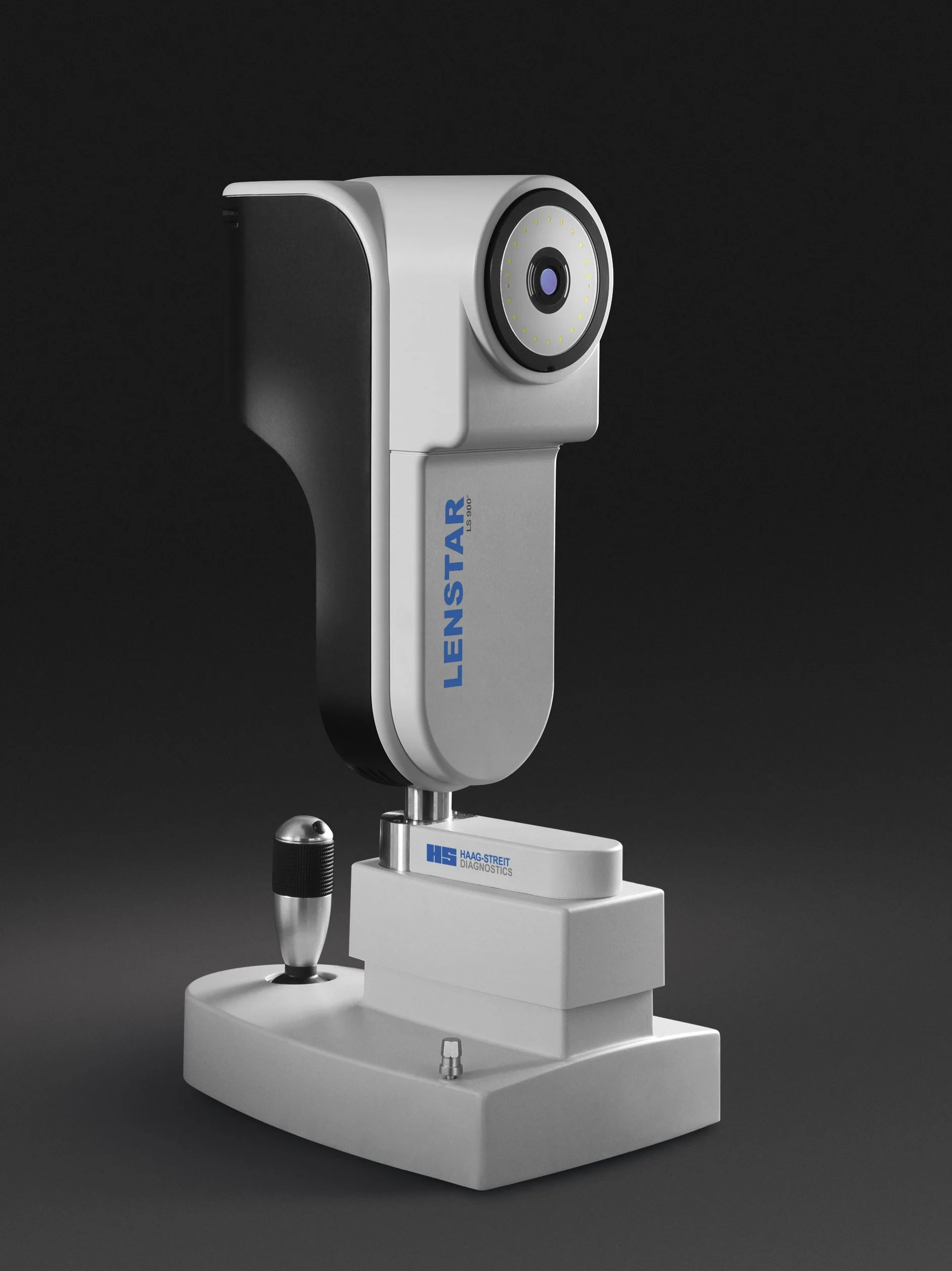
Lenstar Biometry for Myopia Management
The Haag-Streit Lenstar biometer measures the axial length of the eye, a critical measure for assessing the progression of myopia especially in children and adolescents
Home » Technology » Slit-lamp Biomicroscopy and Photography
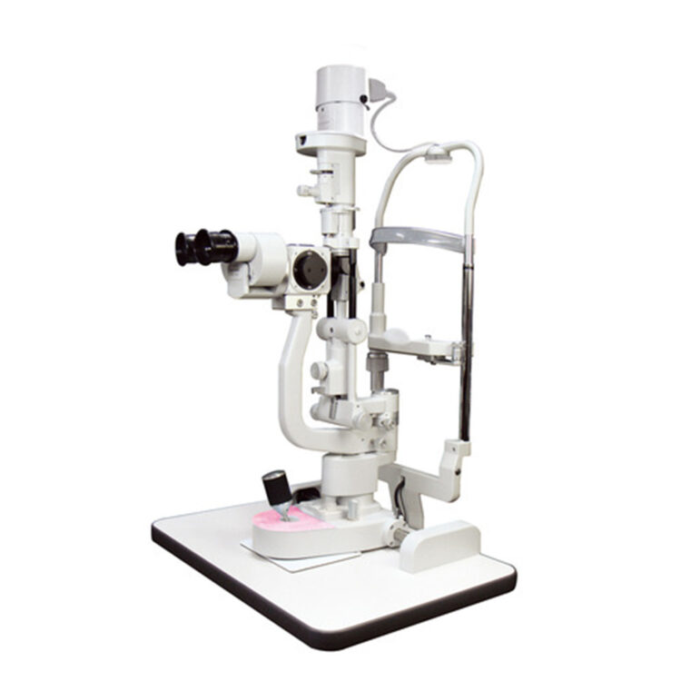
High quality optics, lighting and magnification are essential for seeing subtle signs and changes when examining structures at the front of the eye. These are provided by the biomicroscope, also known as the slit-lamp. Structures that are viewed in detail with the slit-lamp include the transparent cornea and conjunctiva, as well as the coloured iris and white sclera.
Our slit-lamp is fitted with a digital camera that takes both still and video images, and is ideal for recording the appearance of the front of your eye. It is a particularly useful tool for showing the functioning of the meibomian glands and the health of the eyelid margins, both of which are especially important in dry eye disease.

The gonioscope is a fine, smooth mirror that is placed on the cornea, cushioned by thick lubricating fluid. Used in conjunction with the slit-lamp, this enables us to view the anterior chamber angle, where the aqueous humor drains from the front of the eye. Along with images of the angle captured by our optical coherance tomographer (OCT), the slit-lamp and gonioscope are important tools for identifying patients at risk of developing angle-closure glaucoma.
Learn more about the technology that we use at Collins Street Optometrists

The Haag-Streit Lenstar biometer measures the axial length of the eye, a critical measure for assessing the progression of myopia especially in children and adolescents
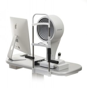
Keratography with the Oculus K5 instrument is an invaluable tool for the assessment, diagnosis and classification of dry eye
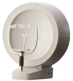
Measurement of visual fields by automated perimetry is an important technique for assessing glaucoma and neurological conditions
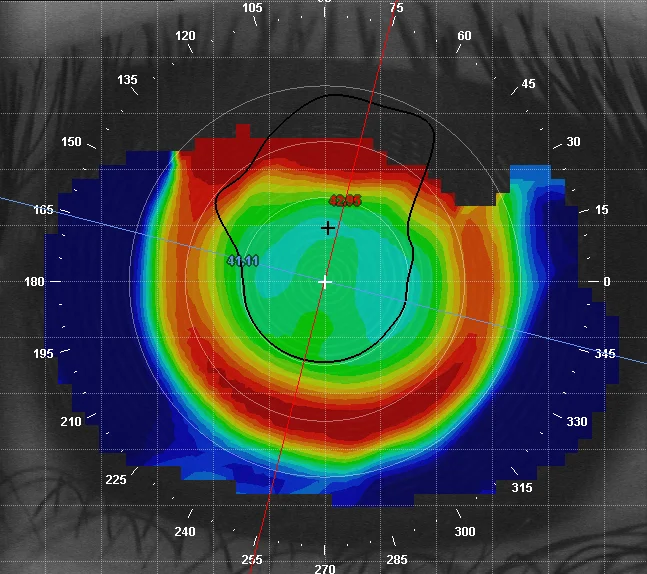
Corneal topography provides a computerised 3-D map of the front surface of the eye, with which we can see the characteristics of its curves and surface irregularities and which is enormously helpful for contact lens fitting
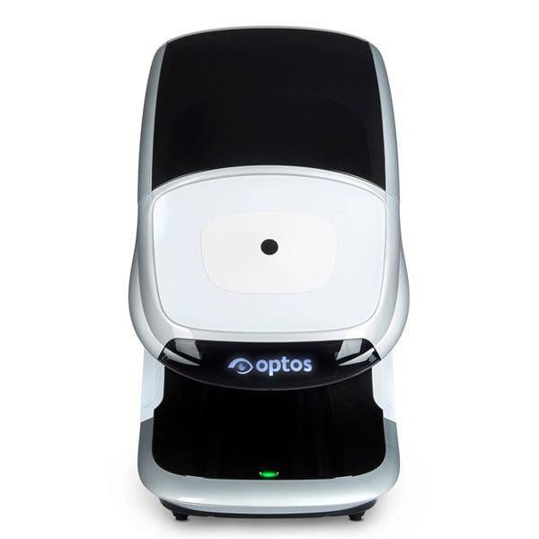
Our Optos Daytona scans the retina with two lasers through a range of 200°, where by comparison, standard digital retinal photography captures only the central 45°
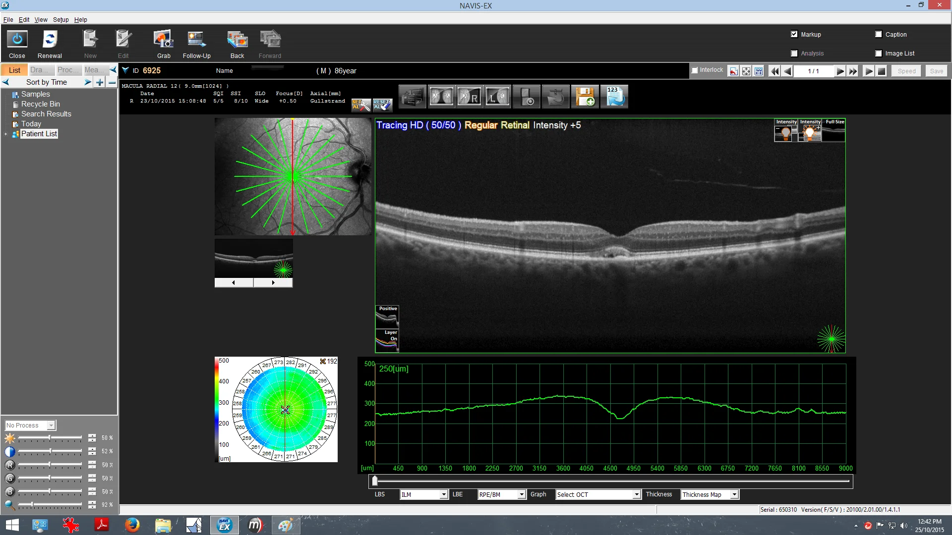
Optical Coherence Tomography is a non-invasive imaging technology used to capture cross-sectional images of the retina and is an essential aid in the diagnosis of retinal disease and glaucoma
Monday - Friday
9AM - 6PM
Saturday - Sunday
CLOSED